Call Us +65 6471 1233

All You Need To Know About Breast Biopsies

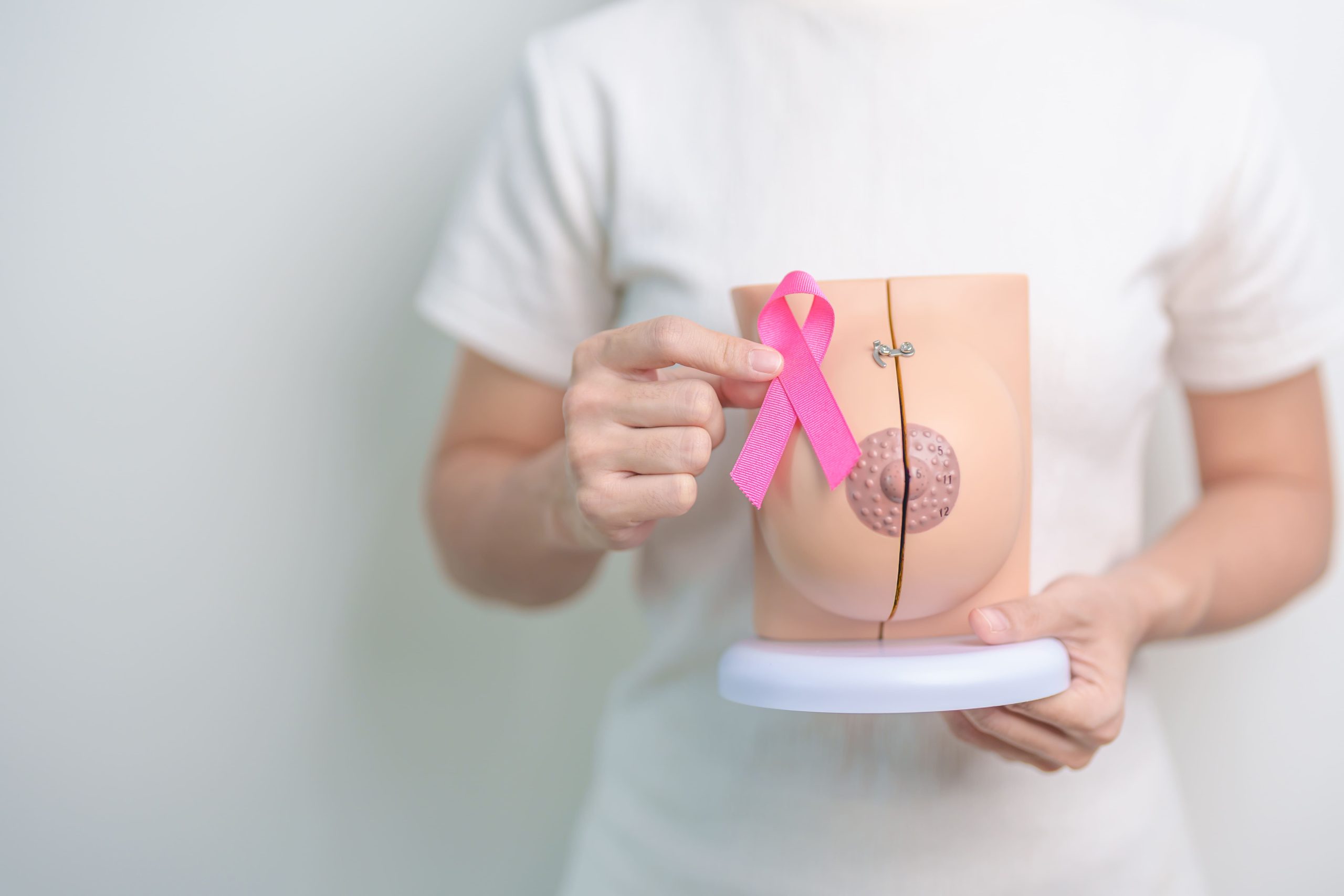
A breast biopsy is a minimally invasive procedure that involves extracting a tissue sample from a suspicious lump or abnormality detected through imaging. It helps determine whether the lesion is benign or cancerous, providing information to guide further treatment. This article aims to provide a comprehensive overview of breast biopsies.

Why Is a Breast Biopsy Recommended?

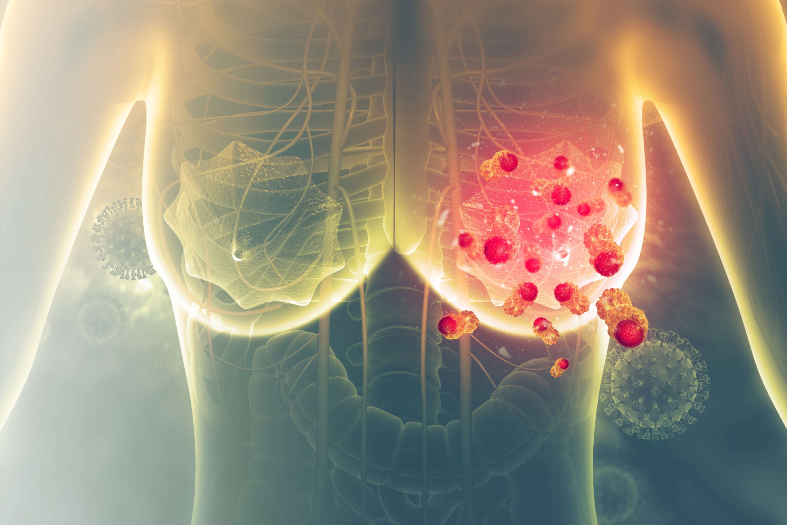
A breast biopsy is recommended when imaging tests, such as a mammogram, ultrasound, or MRI, detect a lesion or abnormality that requires further examination. Because imaging alone cannot always determine if an abnormality is benign (non-cancerous) or malignant (cancerous), the biopsy provides a definitive diagnosis and helps guide appropriate treatment.

Common Reasons for a Breast Biopsy

A breast biopsy is typically recommended when specific signs, symptoms, or imaging findings suggest further examination is necessary. Common indications include:

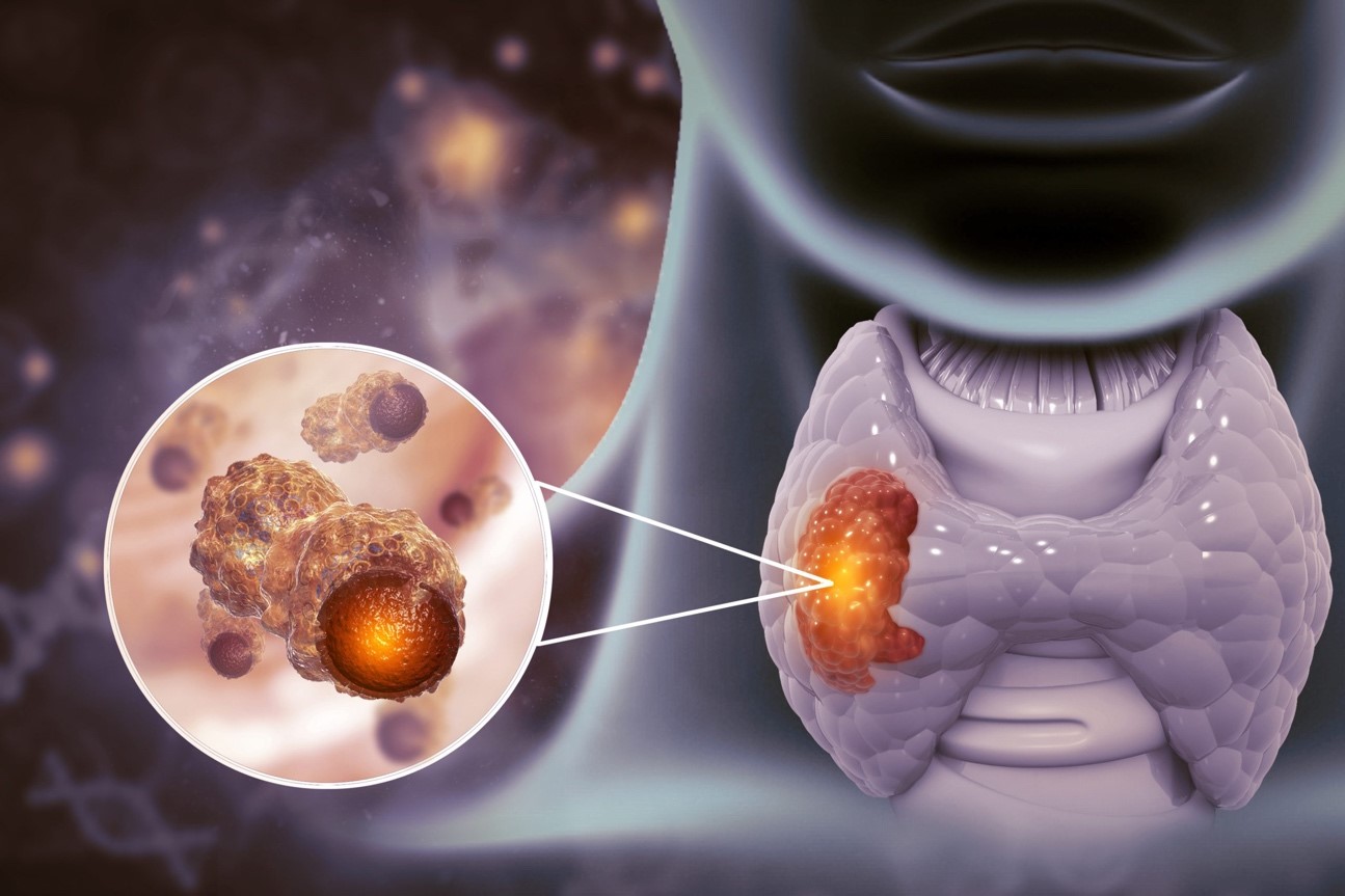
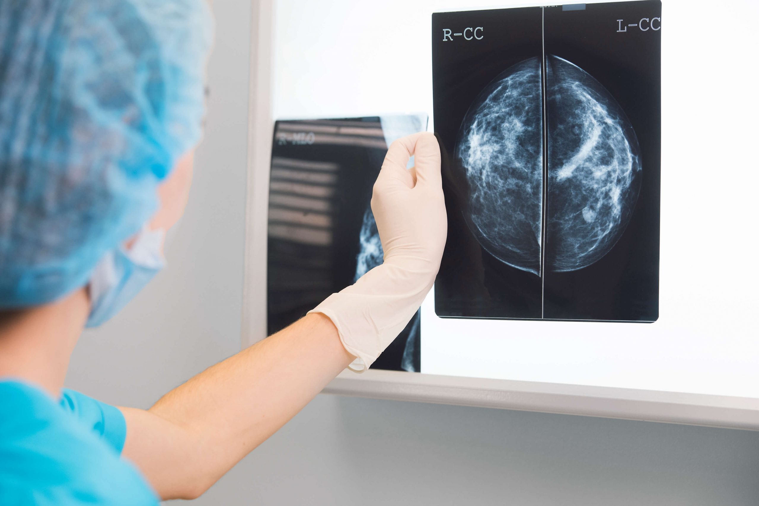
Abnormal Imaging Results: Suspicious findings such as lumps, masses, or calcifications detected through mammograms, ultrasounds, or MRIs may require biopsy confirmation.

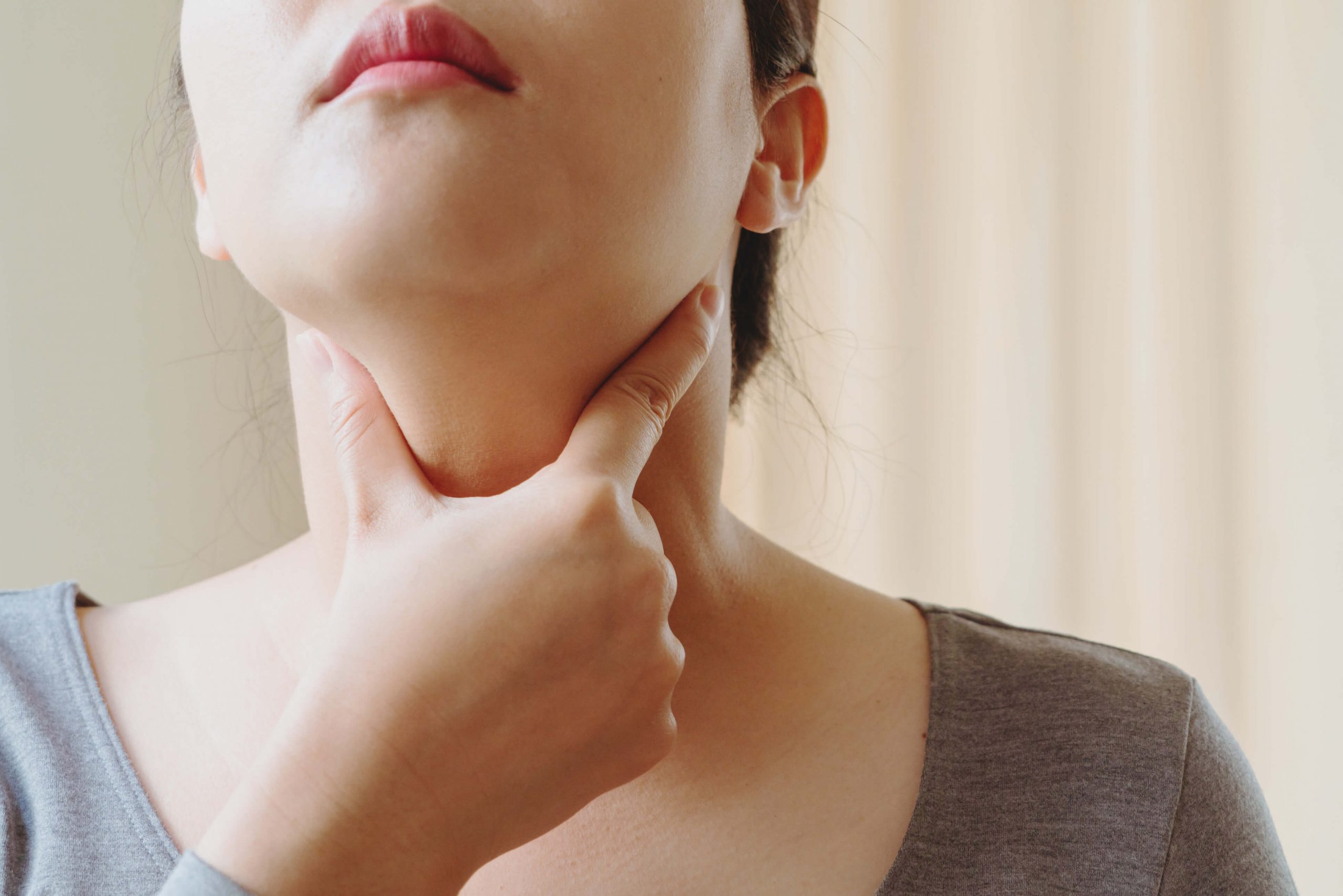
Lumps or Tissue Thickening: A noticeable lump or thickened area identified during a clinical breast exam that feels different from surrounding tissue may indicate the need for further evaluation.

Unexplained Nipple Changes or Discharge: Symptoms such as bloody discharge, inverted nipples, or skin dimpling can suggest an underlying abnormality that requires investigation.

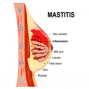
Changes in Breast Texture or Skin: Redness, swelling, scaling, or skin thickening may indicate inflammatory conditions or cancer, prompting the need for a biopsy.

What to Expect During a Breast Biopsy

Preparing for the Procedure

Initial Consultation

Your surgeon will explain why the biopsy is necessary based on your imaging results and what the procedure aims to determine. You will receive a detailed overview of the process, including the type of biopsy performed and what to expect before, during, and after. This is also an opportunity to ask questions, express concerns, and discuss how the results will influence your treatment plan.

Medication Review

Bring a list of all the medications, supplements, and vitamins you take regularly to your appointment. Certain blood-thinning medications, such as aspirin, Plavix, or warfarin, may need to be paused temporarily to minimise the risk of bleeding.

Sedation Option

While most breast biopsies are performed under local anaesthesia, you can discuss the possibility of sedation with your surgeon. Your surgeon will assess your health history and any risks to determine if sedation is suitable.

On the Day of the Procedure

If you are not receiving sedation, you can eat light meals, but avoid heavy food before the procedure. Wash your breasts and armpits with soap and water, and do not apply deodorant.

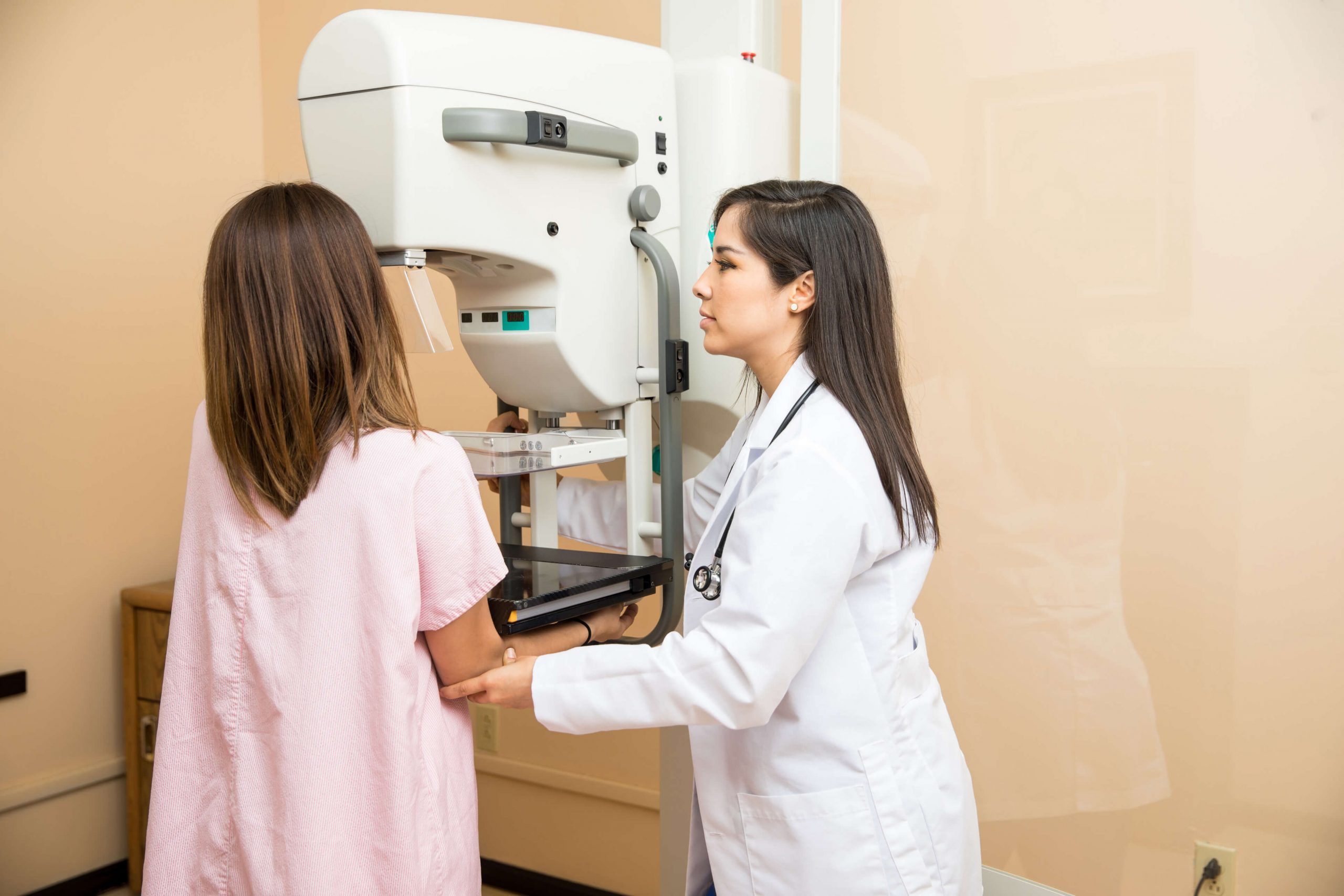
Step-by-Step Procedure

Anaesthesia Administration

Local anaesthesia will be injected to numb the area, ensuring you feel minimal discomfort during the biopsy. If sedation is used, it will be administered before the procedure to help you relax or remain asleep through the process.
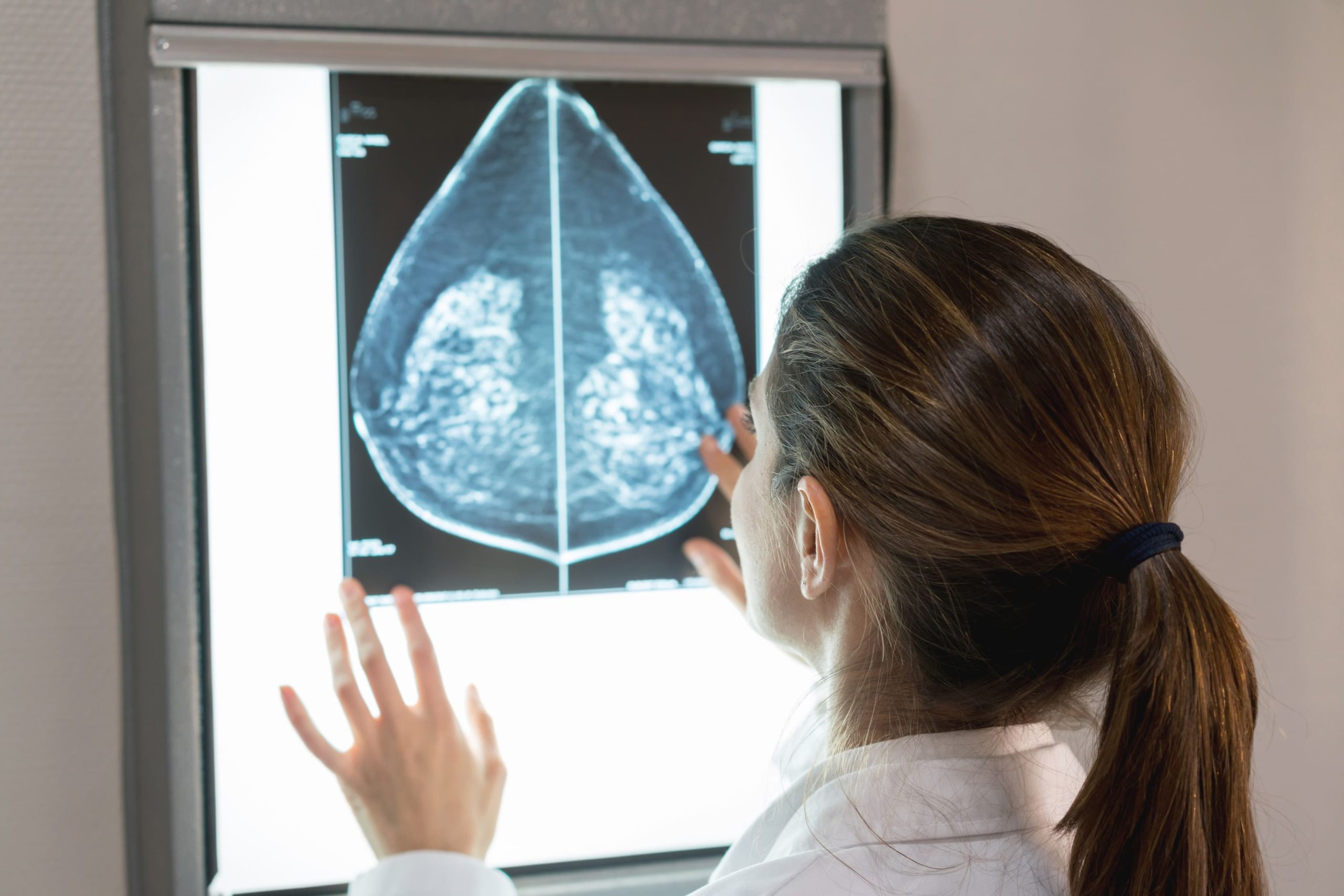
Imaging Guidance

The surgeon will use imaging techniques such as ultrasound, mammography, or MRI to accurately locate the lesion. This guidance ensures the biopsy needle is precisely inserted into the correct area for an effective tissue sample.

Tissue Extraction

A thin biopsy needle will be inserted into the target area to extract a tissue sample for laboratory analysis. If a vacuum-assisted biopsy is used, it collects a larger tissue sample, improving the accuracy of the diagnosis.

Bandage Application

Once the biopsy is complete, the doctor will apply firm pressure to the site to control bleeding and reduce swelling. A bandage or dressing will then cover the area to protect it and promote healing.

Post-Procedure Monitoring

You will be monitored for a short period to ensure there are no immediate complications, such as excessive bleeding or allergic reactions. If sedation was used, you will be observed until the effects wear off.

After Your Breast Biopsy: Care Instructions

Bandage Care

The bandage will remain on the biopsy site to minimise swelling and prevent bleeding. Your surgeon will provide instructions on when and how to remove it safely and how to care for the site.

Pain Management

You may experience some soreness or discomfort after the procedure. Over-the-counter pain relievers, like acetaminophen, are often enough to manage discomfort.

Transportation Arrangements

If you received sedation, the sedative effects may impair your ability to drive or perform tasks that require focus. Make sure someone is available to take you home safely after the procedure.

Follow-Up Appointment

A follow-up appointment is typically scheduled within five days after the biopsy to discuss the results. Your surgeon will explain the findings and, if needed, outline the next steps for your treatment plan.

Possible Risks and Side Effects

Breast biopsies are generally safe, but some patients may experience mild pain, swelling, or bruising at the biopsy site. Infections and bleeding can occasionally occur, though these are uncommon and typically manageable. Following your surgeon’s aftercare instructions helps reduce the risk of complications and ensures proper healing.

Conclusion

A breast biopsy is a necessary diagnostic tool that helps determine whether a lesion is benign or cancerous, guiding the next steps in treatment. With proper preparation and post-procedure care, the process is typically safe and straightforward.

If you have any concerns or need further evaluation, consult Dr Tan Chuan Chien, our Breast Specialist to discuss your options and ensure timely care.

Frequently Asked Questions (FAQs)

Will a breast biopsy spread breast cancer?

When performed by an experienced surgeon, the chance of a breast biopsy spreading cancer is extremely low. The procedure is safe, minimally invasive, and necessary for determining the nature of the lesion and developing an appropriate treatment plan.

Does a breast biopsy remove the entire lesion?

No, a breast biopsy only removes a small sample of the lesion, not the entire lump or mass. This sample is then sent to a laboratory, where it is carefully examined to determine if the lesion is benign or cancerous. The goal is to collect enough tissue to provide a reliable diagnosis without removing the whole lesion.

How long does a breast biopsy procedure take?

The entire breast biopsy procedure typically lasts between 30 and 60 minutes. Additional time is required for preparation, as well as a brief observation period following the procedure. Patients should allow for 1.5 to 2 hours at the clinic.

9 Myths Surrounding Breast Cancer

Breast cancer is one of the most common cancers worldwide, particularly among women, but its prevalence has resulted in the spread of numerous myths. These misconceptions can cause unnecessary fear, prevent early detection, or lead to incorrect assumptions about the disease. In this article, we will investigate and dispel some of the most common myths about breast cancer, allowing you to make more informed decisions about prevention, diagnosis, and treatment.

Myth 1: Only Women Get Breast Cancer

Fact: While breast cancer is far more common in women, men can also develop the disease. Male breast cancer accounts for less than 1% of all breast cancer cases, but the risk remains because men, like women, have breast tissue that can develop cancerous cells. Men must be aware of breast cancer symptoms such as lumps, changes in nipple appearance, or discharge, and seek medical attention if they appear.

Myth 2: Finding a Lump in Your Breast Means You Have Cancer

Fact: Not every lump is cancerous. Many breast lumps are benign and caused by non-cancerous conditions such as cysts, fibroadenomas, or infections. However, all lumps should be checked by a medical professional to rule out cancer. A thorough examination, including a mammogram or biopsy, is required to determine whether a lump is cancerous or benign.

Myth 3: Wearing Underwire Bras Increases Your Risk of Breast Cancer

Fact: This is a long-standing myth that lacks scientific support. Some believe that underwire bras limit lymphatic drainage, resulting in toxin buildup in breast tissue, but this theory has been refuted by numerous studies. The bra you wear has no effect on your risk of developing breast cancer. Breast cancer risk factors are more closely related to genetics, age, hormone levels, and lifestyle.

Myth 4: Breast Implants Increase Your Risk of Breast Cancer

Fact: There is no clear evidence that having breast implants increases the risk of breast cancer. However, a rare form of cancer called breast implant-associated anaplastic large cell lymphoma (BIA-ALCL) has been associated with certain types of implants, particularly textured ones. Women with implants need to continue regular breast cancer screenings and be aware of any changes in their breasts.

Myth 5: Breast cancer is always hereditary

Fact: While having a family history of breast cancer can increase your risk, however, the majority of breast cancer cases are not caused by inherited genetic mutations. Only about 5% to 10% of all breast cancer cases are linked to specific gene mutations, such as BRCA1 or BRCA2. These mutations can significantly raise a person’s risk, but many breast cancer cases arise in individuals with no family history of the disease.

Myth 6: Mammograms Can Cause Breast Cancer

Fact: The small amount of radiation used in mammograms is highly unlikely to cause breast cancer. The benefits of regular mammograms far outweigh the risks, as these screenings can detect cancer at an early stage when it is most treatable. For most women, annual mammograms starting at age 40 (or earlier if you have a high risk) are recommended for early detection.

Myth 7: Breast Cancer Always Requires a Mastectomy

Fact: Not all breast cancer treatments involve removing the entire breast. Mastectomy (the removal of the breast) is one option, but many women can be treated with a lumpectomy, where only the tumour and some surrounding tissue are removed. Radiation therapy, chemotherapy, targeted therapies, and hormone therapies are also available, depending on the type and stage of cancer.

Myth 8: You Can’t Get Breast Cancer if You Lead a Healthy Lifestyle

Fact: While living a healthy lifestyle, including exercising regularly, eating a balanced diet, and avoiding smoking and excessive alcohol, can lower your risk of breast cancer, however, it does not eliminate it. Breast cancer can still occur due to genetic factors, age, and other uncontrollable variables. A healthy lifestyle helps but is not a guarantee against breast cancer.

Myth 9: Breastfeeding Reduces Your Risk of Breast Cancer

Fact: While research shows that breastfeeding can reduce the risk of developing breast cancer, especially if done for an extended period, it is not a guarantee of prevention.

Conclusion

Breast cancer is a complex disease, and knowing the facts is necessary for prevention, early detection, and effective treatment. Misconceptions, like those covered in this article, can lead to confusion, fear, and even delays in seeking appropriate medical care. By debunking these myths, we hope to inspire people to stay informed, prioritise regular screenings, and seek accurate advice from healthcare professionals.

Stay informed and proactive! Schedule your breast cancer screening today and discuss any concerns you may have with Dr Tan Chuan Chien, our Breast Specialist.

What do we need to know about Thyroid Cancer?

Thyroid cancer is more common among women than men. It’s the 8th most common cancer among Singaporean women. It’s also more frequently diagnosed at a younger age than most cancers, with a significant proportion diagnosed before the age of 50 years old.

What is Thyroid cancer?

Cancer is a group of disease that forms when cells become mutated and are able to invade adjacent cells and spread to another organ. Thyroid cancer occurs when normal thyroid cells become mutated to abnormal cells that gains this ability.

How common is it?

Thyroid cancer is more common among women than men. It’s the 8th most common cancer among Singaporean women. It’s also more frequently diagnosed at a younger age than most cancers, with a significant proportion diagnosed before the age of 50 years old.

How does Thyroid cancer present?

Patients with thyroid cancer most often present with a painless lump in the front part of the neck. Other times, they may have a lump at the side of the neck, or notice voice changes or difficulty swallowing.

What are the risk factors for thyroid cancer?

Risk factors are factors that makes a person more likely to develop a certain disease. Risk factors for thyroid cancer includes being female, a personal history of thyroid disease like hypothyroidism, a family history of thyroid cancer, a history of exposure to radiation, and obesity.

How is Thyroid cancer treated?

Treatment depends on the extent of the disease at diagnosis, i.e. The stage of disease.

Doctors may recommend surgery (Total thyroidectomy or hemithyroidectomy) to remove the cancer and affected thyroid lobes, and adjacent lymph nodes as well. Radioactive iodine therapy may also be used after surgery to eliminate any remaining cancerous cells.

Doctors also often prescribe thyroxine (thyroid hormones) to replace what would have been produced by the thyroid gland, and to suppress hormones that stimulate the thyroid, hence suppressing growth of tumour cells.

In certain types of thyroid cancers, targeted therapy and radiotherapy may also be used to treat thyroid cancer.


This article was first published on www.365cps.org.sg on Jan 27, 2023

Telemedicine Guide: How Does It Work?

What is telemedicine?

Telemedicine refers to the use of modern communication technology to deliver healthcare to patients. Traditionally, the patient-doctor interaction requires the patient to be physically present, for a face-to-face consultation. However, as digital technologies have improved and people are more accustomed to using tele-conferencing platforms, particularly since the start of the COVID-19 pandemic, telemedicine has become an increasingly popular alternative to seek medical advice. It allows patients to consult with their doctor remotely at the convenience of their home, minimising unnecessary travelling and reducing the risk of exposure to infection.

How does telemedicine work?

At SOG-CC Tan Breast, Thyroid & General Surgery, we are committed to providing the best care tailor-suited to our patient’s medical needs and comfort levels. We understand that some of our patients may have busy schedules and want to reduce travelling to the hospital, where possible.

If you are eligible, our clinical staff can arrange a telemedicine consultation via the following steps. Upon confirmation of appointment:

  • Clinical staff will email the teleconsultation appointment link to patient
  • To see the doctor, patient can simply click the appointment link at the scheduled appointment time
  • Teleconsultation session will commence through the video-conferencing platform

This gives our patients the flexibility to receive medical advice from their doctor without compromising their busy schedules or other commitments. Read on for a general guide on patients’ eligibility for teleconsultation service.

What conditions are appropriate for telemedicine?

While telemedicine is a great technology that is preferred by many patients, there are some limitations to this form of consultation. Firstly, doctors are not able to perform a physical medical examination on his/her patient through this platform, as it is conducted digitally. Doctors are also not able to perform medical procedures on patients. On the other hand, patients who seek teleconsultations need to be comfortable using these new technologies.

Telemedicine is ideal for patients who have recently consulted their doctor and merely require a quick follow-up consultation or discussion to clarify certain treatment plans. A subsequent face-to-face consultation may be required if the doctor or patient feels that they need further in-person consultation.

Please enquire with our friendly clinic staff if you would like to know more about our teleconsultation service.

Our clinic is listed with the Ministry of Health (MOH) Singapore as a direct  telemedicine service provider.

Breast Cancer Screening In Singapore: What You Need To Know

What Is Breast Cancer?

Breast cancer is the abnormal growth of cancer cells in the breast. These cancerous cells can grow over time and potentially spread to other parts of your body.

According to the Singapore Cancer Society, breast cancer is the most common cancer among Singaporean women, with around 1,000 women diagnosed annually. Furthermore, approximately 1 in 13 Singaporean women will be diagnosed with breast cancer throughout their lifetime.

The common symptoms of breast cancer may include:

  • A lump in the breast or underarm area
  • Changes in the size or shape of the breast (thickening/swelling)
  • Nipple discharge, excluding breast milk
  • Irritation or redness in the breast or nipple area
  • Dimpling of breast skin
  • Pain in the breast or nipple area

Why Is Breast Cancer Screening Important?

Breast cancer screening is crucial as it ​allows doctors to diagnose breast cancer in patients at an early stage, hence enabling simpler treatments and better survival.

Breast cancer usually begins as a precancerous lesion and eventually develops into invasive cancer. It usually begins with changes within the breast that are often not noticeable by the patient. It can be changes that are as small and fine as grains of sands. However, breast screening with scans can detect these changes and enable early diagnosis and treatment of these early breast cancer lesions. Usually, when these lesions are so small, the treatment for breast cancer is much simpler and there is a better chance of survival. Because, breast cancer is not an uncommon disease, hence all women should undergo regular breast screening.

Breast cancer screening as early detection of cancer cells can increase your survival rate with early treatment.

What Is Mammography?

Mammography is an imaging test to examine and detect abnormalities in the breast area using x-rays. It is one of the most common and reliable methods for detecting early breast cancer as it allows the detection of cancerous tumours or lumps that are not large enough to be felt or noticed.

Benefits of Mammography

The advantages of mammography include:

  • Allow detection of early-stage breast cancer before any noticeable symptoms.
  • Significantly increases chances of recovery when cancer is detected early.
  • Simpler treatment if breast cancer detected early hence less side effects and pain.

Risks of Mammography

Similar to most screening methods, there are also risks involved during mammography.

During mammography, your breasts are exposed to a small amount of radiation. As such, there is a risk of radiation due to x-ray procedure. However, as the radiation emitted is of low dosage, there is generally no significant harm to your body.

There is also a possibility of false-positive results, hence, the possibility of over-treatment. ​​For patients with dense breasts, there is an increased risk of false-negative results from the screening mammography, as both the dense breast and cancers may appear white on regular mammography. Unfortunately, we now know that increased breast density is associated with an increased risk of breast cancer. Hence, it is important to discuss this with your Breast Specialist to overcome this via alternate methods of screening, based on your individual risk.

Difference Between 2D and 3D Mammograms

Both 2D and 3D mammograms are largely similar as they use a low-dose x-ray machine to examine the breast area. However, there are still slight differences between them, such as how the images are taken and the manner the results are presented.

A 2D mammogram takes two pictures of each breast to create two images of each breast.

A 3D mammogram takes multiple images of each breast from different angles and shows individual layers of breast tissue. These images are then constructed together to present a 3D view of the breasts. As such, finer details of the breast can be viewed clearly, significantly reducing unnecessary recall and false-negative test results.

What Happens During a Mammogram?

During a mammogram, the x-ray technician will gently compress both your breasts between two transparent plates. Images of each breast will then be taken from different angles (depending on whether you are doing a 2D or 3D mammogram) by special x-ray equipment.

There are generally two types of mammograms, digital and non-digital (film-screen). Both mammograms operate the same way, with the digital method preferred as it can minimise background noise, resulting in clearer images of the breast.

What Are the Alternative Screening Options?

Apart from mammograms, there are also many other additional screening options available. A mammogram is usually sufficient to detect breast cancer. However, there may be instances where further tests may be required in certain scenarios.

These alternative screening options include:

Ultrasound

  • A breast ultrasound scan uses penetrating sound waves (sonography) to look for and determine the solidity of a lump.
  • This is usually done in conjunction with mammography to increase the pick-up rate for abnormalities or suspicious lumps in the breast.

Breast Biopsy

  • A breast biopsy is the next stage of testing and is usually done when your doctor suspects a significant chance of the presence of abnormal cells in your breast.
  • Your doctor will use surgical procedures to sample tissues from suspicious lumps, and these tissue cells will undergo further examination to determine if they are cancerous.

Magnetic Resonance Imaging (MRI)

  • An MRI scan may be recommended if you are under 40 years old and belonging to the high-risk group (family history or presence of BRCA 1 or 2 genes)
  • An MRI scan may also be preferred over mammograms and ultrasounds in cases of silicone injections in your breasts.
  • However, this method is generally not recommended for routine breast screening for normal-risk women.

Clinical Examination

  • A visual inspection of the breast to identify abnormalities or suspicious lumps that can be seen
  • A physical check of the breast to detect unusual lumps

Who Should Be Screened for Breast Cancer and How Often?

According to Singapore national screening guidelines, normal-risk Singaporean women aged 40 and above are recommended to go for a mammogram screening once every year. For those aged 50 and above, a mammogram screening is recommended once every 2 years.

Women in high-risk groups, such as those with a family history of breast cancer or other types of cancer, should see a Breast cancer specialist to discuss their individual risk and may have to commence screening earlier.

You should consult your doctor or breast specialist to discuss your individual risk of breast cancer and the benefits and risks of the various screening options or if you suspect you may have breast cancer due to the detection of abnormalities in your breast.

Conclusion

Breast cancer may be the most common cancer among Singaporean women, but it is treatable if detected early. Early detection through the various screening options is the key to improving the chances of surviving breast cancer.

Contact Dr. Tan Chuan Chien to schedule a breast cancer screening today: https://cctansurgery.com.sg/contact-us/

Lump in Your Breast? What to Do When You Notice One

What Is a Breast Lump?

A breast lump is a bulge or a bump that develops in the breast. Breast lumps can differ in size, appearance and texture. However, they may be too small to be noticeable until detected using screening tools such as mammograms.

Breast Lump vs Normal Breast Tissue

Your breasts are composed of fats, blood vessels, nerves, milk glands, milk ducts, fibrous connective tissues and dense breast tissues. This is a complex anatomy structure, where it can become challenging to differentiate between a breast lump and a normal breast tissue. However, there are still significant distinctions between them.

A breast lump will feel like a discrete distinct and unusual bump that is prominently more solid than the rest of your breast tissues. These lumps also vary in size (from a pea to a golf ball) and texture.

On the other hand, normal breast tissue will have a more consistent spongy and fibrous feel.

Causes & Symptoms of Non-Cancerous Lumps

Most non-cancerous breast lumps and conditions result from a change in breast tissues according to your menstrual cycle and fluctuations in your hormone levels. Other causes may include breast infections or injuries. It is normal for most women to experience changes in their breast tissues throughout their lifetime.

Here are some of the most common causes and symptoms of non-cancerous lumps.

Fibrocystic Changes

It is normal for your hormone levels to fluctuate during your menstrual cycle, resulting in changes in your breast tissues.

These changes are most common in middle-aged women (35 to 50 years old) and are likely to disappear with menopause.

Symptoms of fibrocystic changes include a rubbery or firm texture, a thickening of the breast tissue or a round, fluid-filled cyst.

Fibroadenomas

These are the most common type of benign breast lumps and commonly occur in young women aged 20 to 30 years.

Fibroadenomas are solid, round and rubbery lumps that are movable (akin to a marble ball) and are often painless. These lumps are usually non-cancerous.

Breast Cysts

A breast cyst is a non-cancerous fluid-filled sac in the breast. Breast cysts often develop during parts of your menstruation cycle due to changing hormone levels, and their size and texture often vary throughout the menstrual cycle. These cysts result from the growth of milk ducts and tissues during hormonal release and most commonly affect women aged 35 to 50 years.

Breast cysts are typically tender and smooth under the skin and are filled with liquid. Some of them may be painless, while others are quite painful. Most cysts tend to go away naturally; however, when a breast cyst is particularly large or painful, your doctor may need to carry out a procedure to drain the liquid.

Fat Necrosis

When a breast injury damages fatty tissue, fat will form in lumps. Fat necrosis usually occurs in women with large breasts, particularly in obese women.

These lumps are usually firm and round and are often painless.

Mastitis


Mastitis is an inflammation of the breast tissue due to an infection of the milk duct. It commonly occurs in breastfeeding women, though there are also rare cases of it happening to non-breastfeeding women as well.

This inflammation results in breast pain, swelling, reddening and warmth.

Intraductal papillomas

These lumps are small wart-like growths that develop in the mammary ducts near the nipple and may cause bleeding. Women aged 30 to 50 years are more likely to be affected by this condition.

How Do Cancerous Lumps Feel Like?

Not all cancerous breast lumps look and feel the same. However, there are still some common features of a cancerous lump.

A cancerous lump is typically:

  • Hard
  • Irregularly shaped
  • Immobile (cannot be pushed)
  • Painless
  • Growing over time

It is important to note that a cancerous lump may not necessarily meet any of the above conditions. For example, the lump can be soft and painful.

What to Do If You Noticed a Breast Lump?

Although most breast lumps are benign, some do turn out to be cancerous. If left untreated, a cancerous tumour can spread to other parts of your body. As both cancerous and non-cancerous breast lumps can look or feel the same, it is crucial for you to consult a doctor if you notice any unusual lumps in your breast.

A clinical breast examination is usually the first step of a breast lump diagnosis. During this check, a physical and a visual inspection of the breast will be done to identify abnormalities or suspicious lumps that can be seen or felt. Afterwards, follow-up tests may be recommended by your doctor to evaluate the lump.

How Are Breast Lumps Diagnosed?

There are several methods to evaluate and diagnose a breast lump. Although most breast lumps are non-cancerous, screening and imaging tests are still required to determine and establish the nature of a lump (benign or malignant). The choice of test to be done usually depends on your doctor’s assessment of your situation.

One or more of the following screening and imaging tests may be performed:

  • Mammography. Mammography uses low-dose x-rays to examine and detect abnormalities in the breast. It allows the detection of abnormal changes that may suggest cancerous tumours or lumps that cannot be easily seen or felt. During a mammogram, both your breasts will be gently compressed between two transparent plates. X-ray images of each breast will then be taken from different angles.
  • Ultrasound. A breast ultrasound scan uses penetrating sound waves to determine if a lump is solid or filled with liquid. This may be done in conjunction with mammography to increase the pick-up rate for abnormalities or unusual lumps in the breast.
  • Magnetic Resonance Imaging (MRI). An MRI scan uses magnetic waves to produce detailed images of the interior of your breasts. This method is usually used to detect breast lumps that are not visible with mammography or ultrasound.
  • Breast Biopsy. A breast biopsy is a test usually done when your doctor suspects a significant chance of the presence of abnormal cells in your breast after having done the standard imaging tests. Your doctor will use surgical procedures to remove sample tissues from suspicious lumps, and these tissue cells will undergo further examination to determine if they are cancerous.

Conclusion

Although having a breast lump does not necessarily mean it is cancerous, breast lumps are still the first sign that most breast cancer patients notice. Hence, if you notice a breast lump it is crucial to have it checked as early as possible. Contact our breast surgeon Dr. Tan Chuan Chien, who will help you identify any abnormalities in your breasts and advise you on the next steps. Make an appointment today: https://cctansurgery.com.sg/contact-us/

Mammogram Guide For Women in Singapore

What Is a Mammogram?

A mammogram is an x-ray of the breasts and a screening and diagnostic tool used to examine and detect abnormalities in parts of the breast.

There are generally two types of mammograms — 2-dimensional and 3-dimensional mammograms.

Screening Mammogram

A screening mammogram is a mammogram done on patients with no symptoms or signs to look for early breast cancer.

On the other hand, a diagnostic mammogram is usually performed when you experience any symptoms of breast cancer, such as the development of a lump.

Why Are Mammograms Important?

According to the Singapore Cancer Society, breast cancer is the most common cancer among Singaporean women, with around 1,000 women diagnosed annually. Furthermore, approximately 1 in 13 Singaporean women will be diagnosed with breast cancer throughout their lifetime.

Presently, mammography is the most common and reliable screening method for breast cancer. It allows the detection of cancerous tumours or lumps that cannot be seen or felt. As such, mammograms are crucial in detecting breast cancer early.

In early breast cancer, there may be no noticeable symptoms. Without mammogram screening, you are unlikely to realise if you have breast cancer until there is a lump in your breast that is large enough to be felt or seen. Furthermore, most breast cancer cases are sporadic, meaning that you may be diagnosed with breast cancer even if you have no family history of breast cancer or other risk factors.

As such, it is important to undergo breast cancer screening such as mammograms as early detection of cancer cells can increase your survival rate with early treatment.

Who Is at a Higher Risk of Developing Breast Cancer?

Similar to any type of cancer, there are several risk factors associated with breast cancer. However, possessing risk factors does not mean that you will get breast cancer, as some women may still get breast cancer even without having any. Having a risk factor simply means you have a higher chance of developing breast cancer in your lifetime.

Some of these risk factors include:

  • Gender. Women have a much higher risk of getting breast cancer as compared to men. However, women are not the only ones who can be diagnosed with breast cancer. Men are also susceptible to the disease, with about less than 1% occurrence.
  • Age. The risk of breast cancer increases as you age. Women above 40 years old should have regular breast screening.
  • Family history. Having a first- or second-degree family or relative who has or had breast cancer increases the risk of developing breast cancer.
  • Genetic mutations. Women who have inherited mutated genes such as BRCA1 and BRCA2 have a higher chance of getting breast cancer. In healthy cells, these genes help build proteins that repair damaged DNA. However, mutated versions of the genes can cause abnormal cell development which in turn, can possibly lead to cancer.
  • Hormones. Women who menstruate early (before 12), menopause late (after 55), have their first pregnancy after age 30 or have not been pregnant before are at a higher risk of developing breast cancer. These examples are linked to the female hormone oestrogen, which is known to affect breast tissues.
  • Lifestyle factors. Women who are physically inactive, consume a high-fat diet or indulge in alcohol excessively also have an increased risk of developing breast cancer due to alterations in oestrogen levels. Older women who are obese also have a much higher chance of getting breast cancer. Women who are on Hormone Replacement Therapy (HRT) are also at an increased risk of developing breast cancer.
  • Previous history. Women who have had breast cancer or non-cancerous breast disease are also at a much higher risk of being diagnosed with breast cancer.

How to Prepare for My Mammogram?

Before you go for your mammogram, there are several things you should prepare and take note of to ensure that you have a pleasant experience. Having a proper plan before your mammogram can minimise discomfort, as much as possible.

  • Avoid applying deodorants, body powder, perfumes, or ointments on your breasts or underarms before a mammogram. These substances may appear as white spots during your mammogram, which affects the accuracy of the screening results.
  • Dress comfortably, ideally a two-piece outfit. During the mammogram, you will be asked to remove your top. Hence, wearing something easily removable will be a more convenient choice.
  • Schedule your mammogram preferably a week after your menstrual period. This is to avoid the period of increased breast sensitivity and tenderness, which typically occurs during the menstrual cycle.
  • Consider taking pain relievers or non-steroidal anti-inflammatory drugs such as Ibuprofen an hour before or immediately after the examination to reduce pain.

How Should I Schedule My Mammogram?

According to Singapore national screening guidelines, normal-risk Singaporean women aged 40 and above are recommended to go for a mammogram screening once every year. For those aged 50 and above, a mammogram screening is recommended once every 2 years.

Furthermore, if you belong to the high-risk groups, such as having a family history of breast cancer or other types of cancer, you may be recommended to commence screening earlier.

You should consult your doctor or breast specialist to discuss whether you should undergo a mammogram screening depending on your risk factors and symptoms once you reach the recommended age group.

How Is a Mammogram Done?

During a mammogram, the x-ray technician will gently compress both your breasts between two transparent plates. Images of each breast will then be taken from different angles (depending on whether you are doing a 2D or 3D mammogram) by special x-ray equipment.

You will usually have to stand during the mammogram, and the entire process will take about 15 to 30 minutes, depending on your situation.

What If I Have Breast Implants?

If you have breast implants, you can still undergo a mammogram screening. It is important that you inform your clinic that you have breast implants before scheduling an appointment.

Mammograms for breast implants are slightly different from a standard mammogram. Due to the materials used in implants (silicone or saline), which tend to obstruct the view of breast tissues, it is likely difficult for the x-ray machine to produce a clear view of your breasts.

Hence, your entire mammogram process may require a longer time to complete, as your doctor will need to take additional images of your breasts from different angles (implant displacement views). For these extra images, your implant will be pushed back against your chest, and your breast will be pulled forward over it and then compressed. This allows clearer imaging of the front part of each breast.

What Happens After My Mammogram?

If you have a normal mammogram result, your doctor will likely advise you to continue with future mammograms according to the recommended schedule.

On the other hand, if you have an abnormal mammogram result, you are likely to be called back to undergo follow-up tests such as an additional mammogram view or even a breast ultrasound. However, this does not necessarily mean that you have breast cancer. Most of the time, you may be called back to undergo follow-up tests because the previous mammogram images are inconclusive.

Most abnormal findings on a mammogram are not breast cancer. The changes are usually benign (noncancerous). If your doctor suspects breast cancer, a biopsy may be performed to sample parts of the breast for further testing.

Conclusion

Breast cancer may be the most common cancer among Singaporean women, but it can be treated when detected early. Early detection through mammogram screening is the key to simpler treatments and improving the chances of surviving breast cancer.

Schedule your mammogram with our breast specialist Dr. Tan Chuan Chien: https://cctansurgery.com.sg/contact-us/

Breast Cancer: Symptoms and Early Warning Signs

What Is Breast Cancer?

Breast cancer is the abnormal growth of cancer cells in the breast. These cancerous cells can develop and mutate rapidly, potentially spreading to other parts of your body.

According to the Singapore Cancer Society, breast cancer is the most common cancer among Singaporean women, with around 1,000 women diagnosed annually. Furthermore, approximately 1 in 13 Singaporean women will be diagnosed with breast cancer throughout their lifetime.

Types of Breast Cancer

There are various types of breast cancer, and they can be categorised into two main types: Invasive and non-invasive (in situ).

For invasive cancers, the cancerous cells (malignant) have the ability to spread to adjacent breast tissue and other organs, whereas for non-invasive cancers, the cells do not have this ability.

Some of the most common types of breast cancer include:

  • Invasive Ductal Carcinoma (IDC). IDC is the most common type of breast cancer. It originates from the breast’s milk ducts and slowly invades nearby breast tissues. IDC can also spread to other parts of the body.
  • Invasive Lobular Carcinoma (ILC). ILC begins in the milk glands (lobules) instead of originating from the milk ducts like IDC and slowly invades the other areas of the breast. Similar to IDC, ILC can also spread to other parts of your body.
  • Ductal Carcinoma In Situ (DCIS). DCIS is non-invasive, meaning that the abnormal cells are confined to the breast’s milk ducts and have not spread to nearby breast tissues.
  • Inflammatory Breast Cancer (IBC). IBC is a rare but aggressive form of breast cancer, making up around 1 to 5% of all breast cancer cases. It is classified as Stage 3 breast cancer, where the cancer cells infiltrate the skin and lymph vessels of the breast, blocking the lymph nodes. Apart from developing a lump, IBC can also cause your breasts to swell, turn red and feel very warm.
  • Metastatic Breast Cancer (MBC). MBC is classified as Stage 4 breast cancer, and the cancer cells have spread to other parts of the body such as the lungs, liver, brain or bones.
  • Triple-Negative Breast Cancer (TNBC). TNBC is another uncommon form of breast cancer, affecting only about 10 to 15% of breast cancer patients. For TNBC to be diagnosed, the two hormone receptors (Estrogen and progesterone receptors) and the Her2 receptor are absent from the cancer cells. This type of cancer is more aggressive than the hormone receptor positive breast cancer.

Risk Factors for Breast Cancer

As with any other type of cancer, breast cancer also has its associated risks. However, possessing risk factors does not imply that you will develop breast cancer. Some women may still get breast cancer even though they do not fall under any of the risk categories. Having a risk factor simply means that you have a higher chance of developing breast cancer in your lifetime.

Some of these risk factors include:

  • Gender. Women have a much higher risk of getting breast cancer as compared to men. However, women are not the only ones who can be diagnosed with breast cancer. Men are also susceptible to the disease, with about less than 1% occurrence.
  • Age. The risk of breast cancer increases as you age. Women above 40 years old should have regular breast screening.
  • Family history. Having a first- or second-degree family or relative who has or had breast cancer increases the risk of developing breast cancer.
  • Genetic mutations. Women who have inherited mutated genes such as BRCA1 and BRCA2 have a higher chance of getting breast cancer.
  • Hormones. Women who menstruate early (before 12), menopause late (after 55), have their first pregnancy after age 30 or have not been pregnant before are at a higher risk of developing breast cancer. These examples are linked to the female hormone oestrogen, which is known to affect breast tissues.
  • Lifestyle factors. Women who are physically inactive, consume a high-fat diet or indulge in alcohol excessively also have an increased risk of developing breast cancer due to alterations in oestrogen levels. Older women who are obese also have a much higher chance of getting breast cancer. Women who are on Hormone Replacement Therapy (HRT) are also at an increased risk of developing breast cancer.
  • Previous history. Women who have had breast cancer or non-cancerous breast disease are also at a much higher risk of being diagnosed with breast cancer.

Early Signs of Breast Cancer

In most cases, early breast cancer does not produce any noticeable symptoms. However, there are still several tell-tale indicators of potential breast cancer.

The common symptoms and early signs of breast cancer may include:

  • A lump in the breast or underarm area
  • Changes in the size or shape of the breast or nipple (thickening/swelling)
  • Nipple discharge (clear or bloody)
  • Irritation or redness in the breast or nipple area
  • Dimpling of breast skin
  • Pain in the breast or nipple area

Without screening, you are unlikely to realise if you have breast cancer until there is a significant lump in your breast area that is large enough to be felt or seen. Even so, a lump may not necessarily indicate that you have breast cancer. Breast cancer is often not noticeable, therefore it is important to undergo breast cancer screening as early detection of cancer cells means simpler treatment and less chance of dying from breast cancer.

How Is Breast Cancer Diagnosed?

  • Mammography. Mammography uses low-dose x-rays to examine and detect abnormalities in the breast. It allows the detection of abnormalities that may suggest the presence of cancerous tumours or lumps that cannot be easily seen or felt. During a mammogram, both your breasts will be gently compressed between two transparent plates. X-ray images of each breast will then be taken from different angles.
  • Ultrasound. A breast ultrasound scan uses penetrating sound waves to determine whether a lump is solid or filled with liquid. This is usually done with mammography to increase the detection rate for any abnormalities or unusual lumps in the breast.
  • Magnetic Resonance Imaging (MRI). An MRI scan uses magnetic waves to produce detailed images of the interior of your breasts. This method is usually used to detect breast lumps that are not visible with mammography or ultrasound. It may be recommended if you are under 40 years old and belong to the high-risk group (family history or presence of BRCA 1 or 2 genes). However, this method is generally not recommended for routine breast screening for normal-risk women.
  • Breast Biopsy. A breast biopsy is a test usually done when your doctor suspects a significant chance of the presence of abnormal cells in your breast after having done the standard screening tests. Your doctor will use surgical procedures to remove sample tissues from suspicious areas, and these tissue cells will undergo further examination to determine if they are cancerous.

Conclusion

Breast cancer may be the most common cancer among Singaporean women, but it can be treated when detected early. Early warning signs and symptoms may indicate a more serious issue and could lead to breast cancer. However, these tell-tale signs are often unnoticeable and may not even be present at all.

As such, early detection through the various screening options is the key to simpler treatment and increasing the chance of surviving breast cancer.

Make an appointment with Dr. Tan Chuan Chien for breast cancer screening today: https://cctansurgery.com.sg/contact-us/

5 Common Thyroid Diseases That Affect Women

What Is the Thyroid Gland?

The thyroid gland is a butterfly-shaped organ located in the front of your neck, wrapped around your windpipe (trachea). It is part of the endocrine system responsible for producing hormones, mainly thyroxine (T4) and triiodothyronine (T3). These hormones are essential in many of your body’s processes, helping to regulate your body’s vital functions.

What Does the Thyroid Gland Do?

The thyroid gland plays a crucial role in your endocrine system — releasing and regulating hormones that control your body’s metabolism. This energy is utilised throughout your body to ensure that your body systems are functioning properly. The thyroid gland will typically produce the exact amount of hormones required to keep your body’s metabolism in balance.

Like any other organ in the body, the thyroid can also malfunction, resulting in thyroid-related diseases. In fact, thyroid diseases are relatively common, with women being 10 times more prone to thyroid problems.

Here are the 5 common thyroid diseases that affect women.

1. Goitre

Goitre is a noncancerous type of thyroid disease in which the thyroid gland swells and enlarges. Anyone can develop a goitre regardless of age; however, it is known that women above the age of 40 are more likely at risk.

One or more of the following common symptoms may include:

  • Obvious swelling in your neck
  • Tightness in your neck
  • Difficulty breathing or swallowing
  • Hoarseness of voice
  • Coughing
Causes of Goitre

Iodine deficiency is one of the leading causes of goitres. As iodine is one of the key minerals in the production of thyroid hormones, a lack of iodine in your diet can lead to the development of endemic goitres.

Insufficient thyroid hormones produced can also lead to the enlargement of the thyroid to make up for this shortage, forming a goitre.

Goitre Diagnosis and Treatment

Several tests can be carried out to diagnose a goitre, including the following:

  • Physical examination. Your thyroid specialist may check your neck for swelling or signs of nodules (growth of abnormal tissues).
  • Blood tests can measure thyroid hormone levels to indicate if the thyroid gland is functioning correctly.
  • Ultrasound scan. It is an imaging test that produces images of your neck, your goitre’s size, and any presence of nodules.
  • Nuclear medicine thyroid scan. This is done by injecting a small amount of radioactive material into your veins to produce images of your thyroid on a computer, revealing the condition of your goitre.
  • Biopsy. This involves retrieving several small samples of any thyroid nodules present, which will be sent for further examinations.

The typical treatment methods of goitre are:

  • Medications prescribed depend on whether the thyroid is overactive or underactive.
  • Surgical removal of the goitre.
  • Radioactive iodine treatment to destroy an overactive thyroid.

2. Hyperthyroidism

Hyperthyroidism is a condition in which your thyroid gland produces too much thyroid hormones, and your body cells work faster than usual. This results in a high metabolic rate.

Common symptoms of hyperthyroidism may include the following:

  • Swelling in your neck
  • Rapid and irregular heart rate
  • Diarrhoea
  • Weight loss
  • Fatigue
  • Irregular menstrual cycle
  • Insomnia
  • Elevated blood pressure
Causes of Hyperthyroidism

A variety of conditions can cause hyperthyroidism, including:

  • Grave’s disease, an autoimmune disorder, which causes your immune system’s antibodies to invade and stimulate your thyroid. This will result in your thyroid producing too much hormone.
  • Excess iodine can also cause your thyroid to secrete too much hormone.
  • Thyroiditis, which is an inflammation of your thyroid, will cause stored thyroid hormones to leak out of the thyroid gland.
  • Thyroid nodules, which are benign tumours that develop in your thyroid, may occasionally become overactive and produce too much hormone.
  • Excessive thyroid medication, taken by patients with hypothyroidism (underactive thyroid).
Hyperthyroidism Diagnosis and Treatment

Several tests can be carried out to diagnose hyperthyroidism, including the following:

  • Physical examination. You may be checked for any symptoms such as a rapid pulse or a swollen neck.
  • Cholesterol test. A low cholesterol level may indicate an increased metabolic rate as your body burns cholesterol faster.
  • Blood tests can measure thyroid hormone and thyroid-stimulating hormone (TSH) levels to indicate whether the thyroid gland functions correctly. The pituitary gland, another part of your endocrine system, releases TSH to stimulate the thyroid gland to produce hormones. High thyroid hormone and low TSH levels can indicate an overactive thyroid gland.
  • Radioactive iodine. Your thyroid specialist may administer small amounts of radioactive iodine into the mouth or through an injection to measure the amount of iodine your thyroid gland absorbs, which will reveal whether the thyroid gland is working normally.
  • Imaging tests, such as ultrasound or thyroid scan, can measure the size of your thyroid gland and identify any abnormal growth.

Treatments for hyperthyroidism damages the thyroid gland to reduce the production of thyroid hormones.

  • Medications. Anti-thyroid drugs such as methimazole prevents your thyroid gland from overproducing hormones.
  • Radioactive iodine treatment. A large dose of radioactive iodine may be administered which effectively damages the thyroid gland and the cells that produce thyroid hormones.
  • Surgery to remove part or all of your thyroid can also be done.

3. Hypothyroidism

On the other hand, if too little thyroid hormones are produced, you may develop another condition called hypothyroidism. This is the complete opposite of hyperthyroidism, as your body cells will work slower than expected (underactive).

Some of the common symptoms of hypothyroidism may include the following:

  • Fatigue
  • Constipation
  • Weight gain
  • Swollen neck
  • Irregular menstrual cycle
  • Slow heart rate
  • Depression
  • Muscle aches
Causes of Hypothyroidism

The most common cause of hypothyroidism is Hashimoto’s thyroiditis. It is an autoimmune disorder where your body’s antibodies attack and destroy your thyroid glands, resulting in the underproduction of thyroid hormones.

Over response to hyperthyroidism treatment such as an excess amount of radioactive iodine or anti-thyroid medications may also develop hypothyroidism instead as you may end up lowering thyroid hormone production by too much. Similarly, surgery performed to remove part or all of the thyroid gland may also result in an underactive thyroid as hormone production is significantly reduced.

Hypothyroidism Diagnosis and Treatment

Tests to diagnose hypothyroidism are similar to hyperthyroidism.

They may include:

  • A physical examination to check for symptoms such as a swollen neck or apparent weight gain.
  • Blood tests to measure thyroid hormone and thyroid-stimulating hormone (TSH) levels. Low thyroid hormone and high TSH levels can signal an underactive thyroid gland.
  • Imaging tests, such as ultrasound or thyroid scan, can measure the size of your thyroid gland and identify any abnormal growth.

The typical main treatment of hypothyroidism is to take thyroid hormone medications. These medications replace the shortage of hormones produced by your thyroid gland, ensuring your hormone levels are balanced. However, the amount of thyroid hormone medications to consume must be carefully measured as too much of it may cause hyperthyroidism.

4. Hashimoto’s Thyroiditis

Hashimoto’s thyroiditis is an autoimmune disorder where your immune system’s antibodies attack and destroy your thyroid glands, resulting in the underproduction of thyroid hormones.

Hashimoto’s thyroiditis is different from hypothyroidism as one affects the immune system while the other affects the thyroid gland. It is one of the common possible causes of hypothyroidism. However, if you are suffering from Hashimoto’s thyroiditis, you may not necessarily develop hypothyroidism.

Common symptoms of Hashimoto’s thyroiditis are similar to hypothyroidism.

Causes of Hashimoto’s Thyroiditis

Hashimoto’s thyroiditis occurs when your immune system’s antibodies mistakenly attack and destroy your thyroid glands. The actual reason is largely unknown, but many believe that viruses might play a significant role.

Hashimoto’s Thyroiditis Diagnosis and Treatment

Diagnosing Hashimoto’s thyroiditis is similar to hypothyroidism. Blood tests are performed to measure thyroid hormone and thyroid-stimulating hormone (TSH) levels. Low thyroid hormone and high TSH levels can be signals for Hashimoto’s Thyroiditis. The blood tests can also reveal abnormal antibodies that might be damaging the thyroid.

There is generally no exact treatment for Hashimoto’s Thyroiditis. However, thyroid hormone medications to replace the lost hormones may likely be recommended to raise hormone levels.

5. Thyroid Nodules

Thyroid nodules are abnormal growths that may be solid or liquid-filled, and they tend to develop on or in the thyroid gland.

Thyroid nodules and goitres are not exactly the same. Thyroid nodules are lumps that develop in the thyroid gland, while a goitre is an enlargement of the thyroid gland. A goitre can also develop from the formation of several nodules.
Common symptoms of thyroid nodules are also similar to other types of thyroid diseases.

Causes of Thyroid Nodules

The actual cause of thyroid nodules is unknown, but several medical conditions may result in their formation.

They may include:

  • Thyroiditis, which is the inflammation of the thyroid gland.
  • Iodine deficiency
  • Thyroid adenoma, which is an unusual growth of thyroid tissues
  • Thyroid cancer. Most thyroid nodules are benign, but some do turn out to be cancerous.
Thyroid Nodules Diagnosis and Treatment

During the physical examination, an apparent swelling of your neck may hint the presence of thyroid nodules. Thereafter, imaging tests such as an ultrasound or thyroid scan may be conducted to accurately check for the presence of nodules.

Once a thyroid nodule is detected, blood and thyroid-stimulating hormone (TSH) tests may be performed to measure thyroid hormone levels. A biopsy may also be conducted to determine if the nodule is cancerous.

As thyroid nodules are mostly benign, there is usually no need for treatment. However, if the nodules grow over time and related symptoms appear, treatment may be necessary.

The treatment options for thyroid nodules also depend on whether these nodules result in an overactive (hyperthyroidism) or underactive (hypothyroidism) thyroid. If the thyroid nodules turn out to be cancerous, surgery to remove these nodules will most likely be recommended by a specialist.

Conclusion

Most thyroid diseases such as hyperthyroidism or hypothyroidism cannot be prevented.
However, you can prevent complications by getting diagnosed early and following your doctor’s prescribed treatment.

Schedule a thyroid examination and consultation with our thyroid specialist Dr. Tan Chuan Chien today: https://cctansurgery.com.sg/contact-us/

Breast Lump Removal (Lumpectomy): Read This Before Seeing A Specialist

What Is a Lumpectomy?

Lumpectomy for breast cancer is a surgical procedure to remove cancerous growth or abnormal tissues from the breast. It is also called breast-conserving surgery as only a portion of the breast containing cancerous tissues is removed, thereby preserving the rest of the breasts that are healthy. This is unlike a traditional mastectomy, which removes the entire breast tissue from the breasts.

Why Is a Lumpectomy Done?

Lumpectomy is performed to remove the cancerous growth and prevent it from spreading to other parts of the body. It is also typically less invasive as compared to the entire removal of the breast tissues (mastectomy).

Lumpectomy is done if the following conditions are met:

  • Cancer only affects a portion of your breasts
  • The cancerous tumour is relatively small compared to your breast
  • Ample breast tissues are available to reshape your breast after the removal of the cancerous tissues
  • You are medically fit to undergo surgery and the follow-up radiation treatment

Radiation After a Lumpectomy

Radiation therapy is usually provided after a lumpectomy to remove any remaining cancer cells left in the breast. This significantly minimises the risk of cancer returning (breast cancer recurrence).

This process is generally painless, although minor complications may include skin irritation, redness or peeling.

Who Is Not Suitable for Radiation Therapy?

Radiation therapy is not recommended if you:

  • Are pregnant. Radiation therapy utilises high-energy x-rays, which can harm the fetus. It is recommended that you undergo radiation therapy only after delivery.
  • Previously received radiation treatment in the same breast.
  • Possess medical conditions such as Scleroderma, or other connective tissue diseases, which can increase your risks of radiation side effects.
  • Possess medical conditions such as Scleroderma, or other connective tissue diseases, which can increase your risks of radiation side effects.

How Will Your Breasts Change After a Lumpectomy?

Your breasts will likely change in terms of appearance and texture after a lumpectomy. This is due to the removal of cancerous breast tissues, which will decrease the size of your breasts. Furthermore, if you have a larger tumour than breast size ratio, your breasts may become asymmetrical.

However, you may opt for breast reconstruction or other cosmetic surgeries such as liposuction and oncoplastic breast surgery either during your lumpectomy or later. This is usually done to maintain a more natural appearance of your breast and also to restore breast symmetry.

What Happens During a Lumpectomy?

During a lumpectomy, your doctor will first administer anaesthesia before making incisions on your breasts. They will then locate the cancerous tumour and carefully remove it, together with a rim of some healthy breast tissues surrounding the tumour. This is done to ensure that no cancer cells are left behind. The tumour is then sent for testing.
Additionally, your surgeon may also remove any lymph nodes from your underarm to check if the cancer has spread.

Lastly, your surgeon will likely place small marking clips into the lumpectomy site to aid your radiologist to identify the area required for radiation therapy. The breast tissue is then sewn back together with stitches and then bandaged.

What Are Possible Complications?

Similar to any other surgery, there are also possible complications after a lumpectomy.

They may include:

  • Bleeding
  • Infection
  • Pain
  • Temporary swelling
  • Numbness due to the possibility of nerves being affected during tumour removal
  • Changes in the size and appearance of your breasts, especially if a large portion is removed.
  • Formation of scars at the surgical site

How to Prepare for a Lumpectomy?

Before your lumpectomy, you will likely have several appointments scheduled with your breast surgeon. They may perform physical examinations or imaging tests such as mammography to determine the shape and size of the tumour.

They may also ask about the type of medications you are currently taking or any allergies that you may have.

Some of the things that your doctor may further recommend include:

  • Avoid taking blood-thinning medications such as Aspirin up to a week before the surgery to reduce your risk of bleeding after the surgery.
  • Avoid eating or drinking 8 to 12 hours before the surgery.

It is also ideal that you bring along a family member or a close friend during the day of your surgery to provide support and help you take down crucial information relating to the surgery or post-surgery.

What to Expect After a Lumpectomy?

Lumpectomy surgery can be done as a day surgery procedure, meaning you can generally go home after the surgery.

After the surgery, you will be transferred to the recovery room, where you will be observed and monitored until your anaesthesia wears off. Depending on your situation, you may have a small tube inserted in your chest at the surgical site, where it will be attached to small bags for liquid to drain. Your doctor will likely remove this small tube within a week or so. During this period, you will have to empty the drain several times a day.

Following that, your doctor will likely advise you on how to care for yourself at home during the recovery period, which may include:

  • Avoiding any strenuous activities and ensuring that you get sufficient rest.
  • Taking any pain medications as prescribed and recommended by your doctor.
  • Wearing a bra with good support to minimise movement as much as possible to prevent additional pain.

If radiation therapy is required, it will typically commence within a few weeks after lumpectomy.

How Long Does It Take to Recover?

While most patients are able to go home the day after their surgery, the recovery period from a lumpectomy surgery varies for each person. The recovery process depends on the severity of your cancer, how much breast tissue is removed during the surgery, your general health, age and many other factors.

For 1 to 2 days after the surgery, you will likely feel fatigued and experience minor pain. Swelling and bruising may also last for several weeks.

It will generally take from about 2 to 3 weeks to a month to recover fully from the surgery. However, it depends on whether additional surgery had been performed, or further radiation therapy, as these additional treatments will likely lengthen the recovery time.

When Should You See Your Surgeon?

It is not the end even after you fully recover. You should schedule follow-up appointments with your doctor to review your condition.

Contact your doctor to seek for immediate medical attention if you experience:

  • A lump that does not disappear even after your menstrual period
  • Any changes in the size or appearance of your breasts
  • Blood or clear fluid discharge from your breasts
  • Bleeding, especially at your breast area
  • Breathing difficulties
  • Dizziness
  • Chest pain or tightness
  • Prolonged pain even after taking painkillers

Conclusion

Most of the time, the intent of breast cancer surgery is to remove the cancer completely. However, there is still a risk of breast cancer recurrence in some cases. As such, it is crucial to go for all of your follow-up appointments as scheduled with your doctor, and constantly take note of any abnormalities or symptoms.

It is also important to seek treatment early. Early detection and treatment of breast cancer significantly increases survival rate and a chance of recovery.

Consult our breast surgeon, Dr. Tan Chuan Chien, to know more about lumpectomy surgery, or discuss any concerns you may face: https://cctansurgery.com.sg/contact-us/